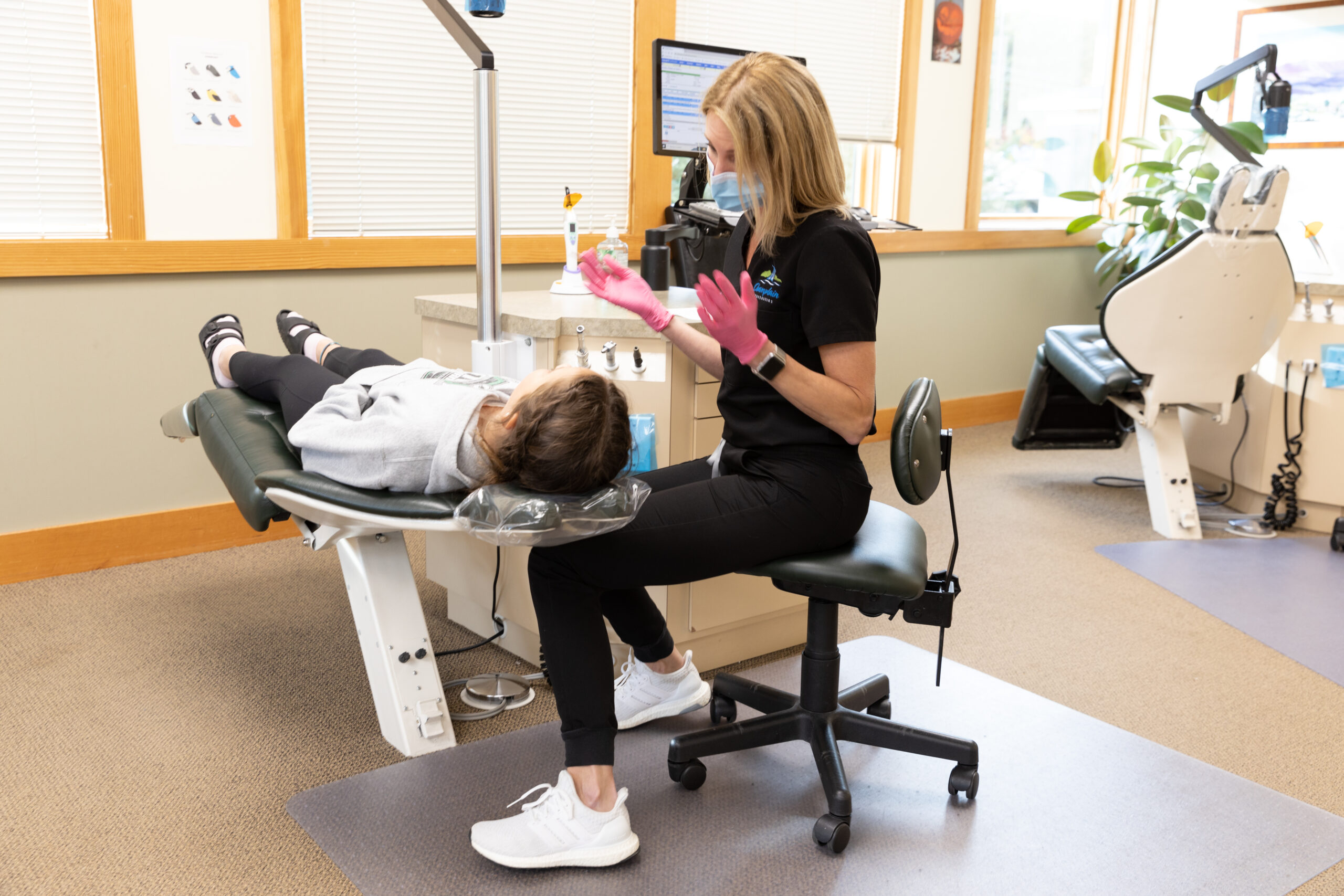
We use many tools at Champlain Orthodontics to create a unique treatment plan, x-rays being one of them. You may be curious about how and why orthodontists use x-rays in the office. Their science lets us see what is going on internally within your teeth and jaw and gives us a better idea of how to help you reach your smile goals! Read on to learn more about everything x-rays provide us with and what to expect when you get one yourself.
The Foundations of X-Rays
Commonly referred to as dental radiography, x-rays are a diagnostic tool used to examine the internal happenings of your body. In the case of orthodontics, we use them to learn more about the alignment of your teeth and jaw, your bone structure, and the roots of your teeth. Electromagnetic waves are emitted to take these images, using minimal radiation to reveal your bone and tissue.
How Dental and Orthodontics X-Rays Contrast
You most likely have received an x-ray from your dentist around once a year. Should your last one have been taken six months ago or earlier, we are able to use it for reference. Otherwise, we may need to take another. Dental x-rays focus on oral concerns, including tooth decay, your roots, teeth growth, and cavities.
While all of this is important for Dr. Ryan and Dr. Eaton to consider in your treatment plan, they could need additional information included in an orthodontic x-ray, as mentioned above, one of the most important being details on your current alignment.
What Information Do They Provide?
This non-invasive process provides our team with a wealth of information necessary to figure out what orthodontic treatment is required and the most effective way of getting there. We also use them to monitor your progress throughout orthodontic care to ensure your teeth are moving in the correct direction.
Is There Any Risk Involved?
There is very minimal risk involved in receiving an x-ray. The benefits outweigh the risks. We are able to learn information from this process to prepare your unique treatment plan. Because they only take a few minutes, you are exposed to minuscule amounts of radiation that are additionally combated with the help of safety measures such as wearing a lead apron.
X-rays are a common tool used in many medical fields and are well-researched to protect patients as best as possible so we can get the details we need to offer you expert care.
What Are The Types Of X-Rays Taken?
Panoramic
The first type of x-ray we take is called panoramic. Similar to a view of a panoramic photo taken on your phone, it captures a wide angle of your teeth, jaw, alignment, and temporomandibular, or jaw, joints. This helps us gain a better grip on your current development so we can use this information to create a comprehensive care plan.
Cephalometric
This second x-ray available is known as the cephalometric. More simply referred to as the orthodontic x-ray, this radiograph allows us to see an exceptionally detailed view of your upper and lower jaw. The results show us how they interact with one another and understand the relationship of your facial bones. This intricate perspective helps us understand how your teeth typically work, information that is vital in preparing for your treatment.

Explaining the Procedure
Safety Precautions and Set Up
The first step in preparing for your x-ray is providing you with safety gear. Even with minimal risk, we take every precaution to keep you safe by placing a lead apron over your head to prevent radiation from reaching areas of your body that do not participate in the x-ray. You will also be asked to remove metal objects from yourself, such as jewelry, hearing aids, hair clips, and glasses, to prevent a warped image.
Guiding You Into Position
Next, we will help you move into the correct position so our machinery has a perfect view of your teeth and jaw. Our team will double-check that you are set before beginning the machine, so you will only be done in a few minutes.
Capturing The X-Ray
Now is the easy part! All you need to do is stay in position while our treatment coordinator sets up the system to take the necessary images during this painless process. A focused beam of radiation is emitted toward your teeth and jaw or any other area we need to capture pictures of. Our treatment coordinator will let you know when the process is complete so you can go back to a normal position.
Analyzing Your Results
Just like that, your x-ray is finished. Thanks to the advancements in digital x-rays, the entire procedure will take fewer than five minutes. You can then peek at your results before they are printed and prepared for Dr. Ryan and Dr. Eaton to offer their expert thoughts.
Keeping Track Of Your Progress
Pre-Treatment
Following the procedure detailed above, Champlain Orthodontics will take an x-ray of your teeth before officially beginning your treatment, most likely during your free consultation. Our orthodontists will examine the results so we can figure out the current positioning of your teeth and determine where we want them to be at the end of treatment.
During Treatment
Only in rare cases do we suggest getting an x-ray in the middle of treatment, but you can expect it during the final phase or the last six to nine months so we can identify areas of your teeth that still need fine adjustments to reach the ideal positioning.
Post-Treatment
To evaluate if the results we can physically see match the work done to your bones and jaw inside, an x-ray may be taken to ensure your treatment is officially complete. We also use these results to help determine how to keep your new smile intact long-term.

Your Free Consultation Awaits!
With offices in St. Albans and Williston, Champlain Orthodontics is proud to provide expert treatment to the community and provide lifetime smiles. If you have any questions about x-rays or orthodontic treatment, please don’t hesitate to contact our front desk. Schedule your free consultation with us today!